![]()
A Database of Polysachharide 3D structures

| Home | |||||||
| About | |||||||
| User Guide | |||||||
| > Xylan Structures | |||||||
| Methods | |||||||
| References | |||||||
| Wiki | |||||||
| Contact us | |||||||
|
** External Links **
|
||||||
|
|||||||
Discover Polysaccharides |
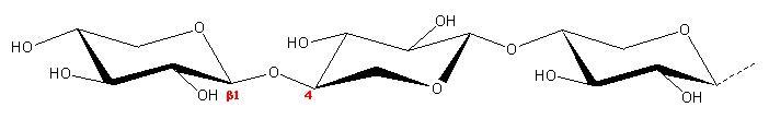
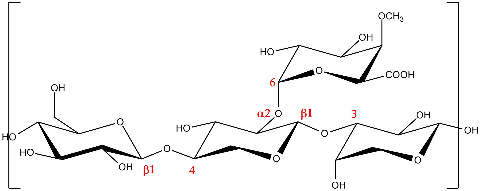
Xylans......................................................................................... Xylans constitute a family of polysaccharides which are present in the main groups of plants, the angiosperms. They can be divided into the group of (glucurono-) arabinoxylans and glucuronoxylans. In the cell walls of monocotyles (grasses and cereals) xylan consist of linear chains of β-(1→4)-linked D-xylopyranosyl residues, which can be substituted with α-L-arabinifuranosyl at the 2-O and 3-O positions and α-D-glucopyranosyl uronic acid or its 4-O-methyl derivative at the 2-O positions. A variety of dimeric or trimeric side chains have been identified as minor substituents. They can be composed of arabinose only, or can include xylose and galactose residues. As with other plant polysaccharides, the source from which the xylan is extracted strongly determines the specific features with respect to the type, the amount, position and distribution of substituents over the xylan backbone. Within one plant source, different populations of xylan may occur. In contrast to the xylan from monocotyles, xylan from dicotyles (hard woods, grass and woody plants) is an O-acetylated 4-O-methyl α-D-glucuronoxylan, almost without any arabinose substitution. On average, every tenths xyloxyl residue carries a a-4-O-methyl glucuronic acid residue substituted at the 2-O-position. O-acetyl substituents can be located at the 2–O and/or 3-O-positions of the xylosyl residues present.
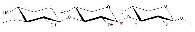
a. b. c. Fig.1 Schematic representation of the repeating unit of (a) β-(1→4)-D-Xylan, (b) β-(1→3)-D-Xylan, (c) Xylan having both β-D-(1→-4) and β-D-(1→3) linkages
There has been some structural analysis performed from an X-ray diffraction pattern of low quality
|