![]()
A Database of Polysachharide 3D structures

| Home | |||||||
| About | |||||||
| User Guide | |||||||
| > Nigeran Structure | |||||||
| Methods | |||||||
| References | |||||||
| Wiki | |||||||
| Contact us | |||||||
** External Links **
|
|||||||
|
|||||||
Discover Polysaccharides |
Nigeran
......................................................................................... Nigeran is a polysaccharide found in the cell wall of lower fungi. In certain Aspergillus and Penicillium spp. Nigeran was first isolated from Penicillium expansum and Aspergillus niger
Nigeran is part of the hyphal cell wall, where it can contribute up to 40% of the cell dry weight. Nigeran chains occupy several domains or location on the hyphal wall
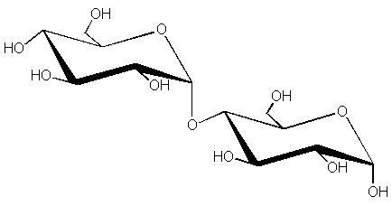
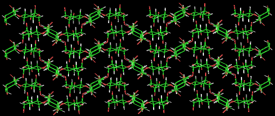
Chemically, nigeran is a regular linear polysaccharide made of α-D-glucopyranose units alternatively linked (1→3) and (1→4)
Fig. 1 Diagrammatic representation of the nigeran structure
The degree of polymerization can be as large as 3000. Nigeran is unusual in its propensity to accommodate or lose water molecules in reversible transition that occur in the crystalline state. A method for growing single crystals of this polysaccharide has been developed (dimension of the single crystals). The X-ray diffraction pattern recorded from a mat of these single crystals indicated a fiber repeat having two-fold symmetry with a repeat of 14.62 Å. With proper control of relative humidity and temperature, nigeran can be induced to assume different packing modes in its crystalline state. Using electron diffraction methods on these single crystals it has been demonstrated
The crystal structure of anhydrous nigeran was determined by using a combined electron diffraction, X-ray diffraction and molecular modelling analysis
a.
b.
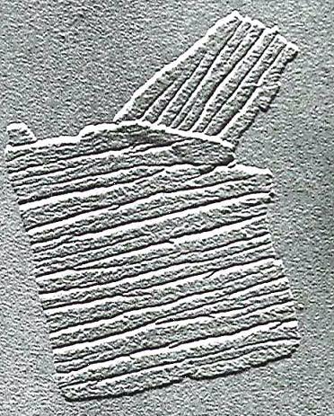
c. Fig. 2 Nigeran (anhydrous) : (a.) Electron micrograph. (b.) Electron diffraction pattern. (c.) Fiber diffraction pattern.
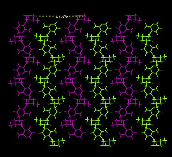
The polysaccharide chain is a two fold helix stabilized by an intrachain hydrogen bond between contiguous α (1→4) glucose residues. Two such polysaccharide chains pack with anti-parallel polarity and the twofold screw axis coincides with the macromolecular axis. A dense network of hydrogen bonds holds the chains together in the crystal.
Fig. 3 Nigeran chains : (a.) Along the X-axis (b.) Along the Y-axis
Nigeran provides an interesting illustration of the mechanism of interaction of hydrolytic enzymes acting on polysaccharide crystals. When treated with an endo-mycodextranase the crystals tend to break into “jigjaw” fragments in which the relation to the original morphology is still visible. Nigeran crystals are composed of folded chains, as evidence from their growth form high molecular-weight material. They occur as a mosaic of tightly folded blocks, linked together by loosely folded, and connecting nigeran chains.
Fig. 4 Schematic representation of the folded chains making up the crystal of nigeran. When performed at ambient temperature, the enzymatic digestion operates on the loose folds, leaving the components of the mosaic unaltered. This indicates that the erosion starts at the crystalline platelet surface, where long and flexible stems are accessible to the enzyme active sites. The folded lamellar morphology of nigeran in its crystalline region may fulfill a protective function because it protects the polysaccharide (and thus the wall) from enzymatic attack.
|