![]()
A Database of Polysachharide 3D structures

| Home | |||||||
| About | |||||||
| User Guide | |||||||
| >Mannan structures | |||||||
| Methods | |||||||
| References | |||||||
| Wiki | |||||||
| Contact us | |||||||
** External Links **
|
|||||||
|
|||||||
Discover Polysaccharides |
Mannan
......................................................................................... Mannan, is a polysaccharide made of β-D-mannose units linked 1→4, that can be obtained as a pure homopolysaccharide of the endosperm of certain plants, notably ivory nuts: Phyelephas macrocarpa. It is also found in the walls of algae belonging to the families Codiacea and Dasycladaceae.
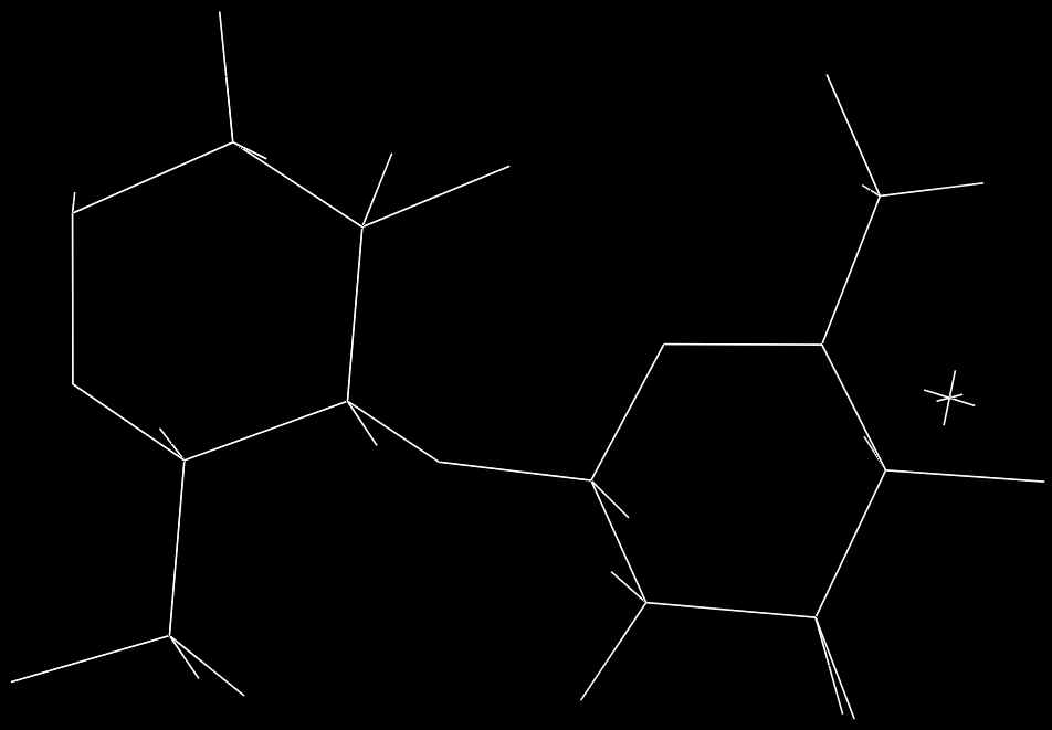
Fig. 1 Schematic representation of the repeating unit of Mannan (X-ray diffraction)
Following preliminary X-ray studies
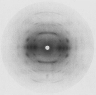
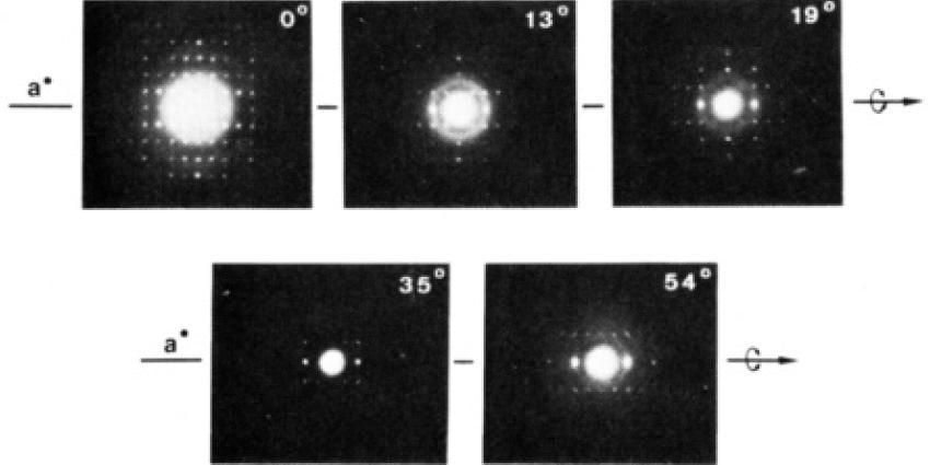
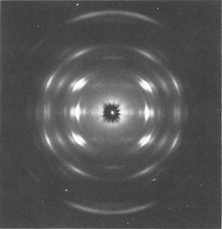
a. b. Fig. 2 (a.) X-ray diffraction diagram form mannan I showing the 2-fold helix symmetry and a c fiber repeat of 10.27Å (b.) Electron diffractogram of Mannan I
In the later case, the intensities were measured from diffraction patterns produced by tilted specimens; several zones were collected, digitized and reduced to integral intensities. Both studies agree and propose a 3D structure which is made up of extended two fold helices stabilized by intramolecular hydrogen bonds in agreement with the stereochemical features of one of its model compounds : mannotriose. The chain is stabilized by two intramolecular hydrogen bonds, O3H…O5 (2.58 Å) and O6H…O3 (3.0 Å).
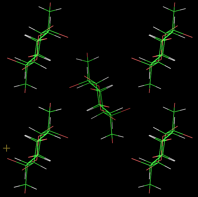
Fig. 3 Schematic representation of the crystalline conformation of Mannan I
Within the orthorhombic unit cell, the two chains are related by crystallographic screw axes perpendicular to the chain direction and, hence, are antiparallel. The packing consists of alternatively “up” and “down” chains along the apparent growth plane. The association of adjacent chains is stabilized by intermolecular hydrogen bonds.
X-ray analysis
Fig. 4 X-ray diffraction pattern of Mannan α 1→3
Xylomannans are extracted from red seaweed Nothogenia fastigiata. Potential activities: anti-viral, helpes simplex virus type 1 and 2 in vitro activity, inhibits replication of herpes simplex (HVS) 1 and 2; human cylcomegalovirud (HCLV), respiratory syncytial virus (RSV), influenza A and B, Junin virus, Tacaribe, Simina virus. |