Inulin
.........................................................................................
Introduction
Inulin is a linear plant polysaccharide belonging to the family of fructans which, after starch, are the most abundant non-structural polysaccharide found in Nature. Inulin is available in large quantities from the roots of chicory. It is also abundant in the tubers of Jerusalem artichokes as well as those of dahlias and can be found in small amount in other plant species. As human lack the glyochydrolase inulinase, inulin is not metabolized directly. Nevertheless, since inulin can be utilized by micro-organism living in the large intestine, it is considered important to the human diet. Acid hydrolysis of inulin yields D-fructose and fructose containing oligomers with great potential as sweeteners. Complementary to its food applications, inulin is also used in the biomedical field as a diagnostic agent for renal functions.
Inulin is poly(2→1)-β-D-fructofuranan ending by an α-D-(1→2) glucopyranose ring. There are reports of inulin with a degree of polymerization as high as 100 as extracted from some plants. In some cases, high molecular weight inulin has been reported to occur in fungi and bacteria.
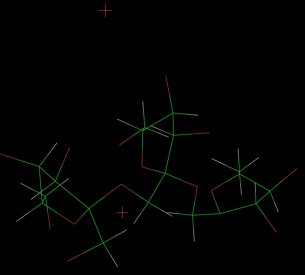
Fig. 1 Schematic representation of the repeating unit of inulin hemihydrate
Normally the molecular weight of inulin is too low to be amenable to an X-ray fiber diffraction investigation. Sample having with a low molecular weight can be crystallized readily from aqueous solution, but not from anhydrous media. It can be crystallized easily in the form of lamellar single crystals having dimensions amenable to structural investigations from electron diffraction data  .
.
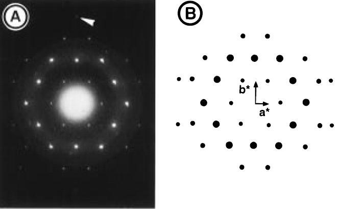
Fig. 2 Comparison of the observed (A) and simulated (B)
electron diffraction patterns for the base plane (hk0) of inulin
hemihydrate. The arrow points toward the 040 reflection that
is weak in the observed diffractogram but absent in the
simulated pattern.
The crystal and molecular structures of the hemihydrate and monohydrate forms of inulin (extracted from chicory) have been established from electron diffraction data and application of the a constrained linked-atom least squares refinement procedure . In the hemihydrate form, there is half a water molecule per fructosyl residue and the crystals belong to the orthorhombic P212121 space group with cell parameters a=16.70 Å, b = 9.65 Å and c = 14.4 Å (chain axis). The structure is based on two antiparallel six-fold helices centered on the two-fold screw axis intersecting the base plane of the orthorhombic unit cell; the six water molecules are distributed over eight sites in the unit cell.
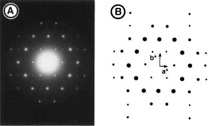
Fig. 3 Comparison of the observed (A) and simulated (B)
electron diffraction pattern for the base plane (hk0) of inulin monohydrate.
The monohydrate form of inulin is obtained when the crystals are kept in a moist environment; there is one water molecule per fructosyl residue. While the unit cell dimensions are almost unaltered (a=16.70 Å, b = 9.80 Å and c = 14.7 Å (chain axis)) substantial differences occur in electron diffraction intensities. Elucidation of the crystalline arrangement, along with simulated diffraction patterns of this structure indicate that the differences with the hemihydrates form could be accounted for only by introducing extra water molecules into the gaps of the crystalline structure without major modification of the inulin conformation.
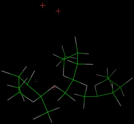
Fig. 4 Three dimensional representation of the hemihydrates form of inulin monohydrate.
It remains to be seen whether the hydration pattern encountered in inulin has any specific ecological and biological significance.