![]()
A Database of Polysachharide 3D structures

| Home | |||||||
| About | |||||||
| User Guide | |||||||
| > Curdlan Structures | |||||||
| Methods | |||||||
| References | |||||||
| Wiki | |||||||
| Contact us | |||||||
** External Links **
|
|||||||
|
|||||||
Discover Polysaccharides |
Curdlans
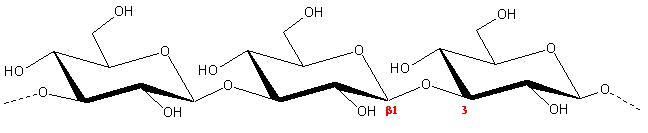
......................................................................................... The generic term « curdlan » refers to extracellular microbial polysaccharides having (1→3)-linked β-D-glucose residue as their repeating units.
Fig. 1 Schematic representation of the repeating unit of Curdlan
It concerns also, a branched β-(1→3)-glucans with a few β-(1→6) glucose units groups among which is scleroglucan, lentinan, schizophyllan,…. These polysaccharides often form a triple helical conformation with a tendency to give a physical gel. Scleroglucan, lentinan, schizophyllan,…. all share similar structures and conformations. Their solubility increases in alkaline conditions, but an irreversible helix-coil transition occurs over pH > 12. Many of the polysaccharides belonging to this family are claimed to have anti-tumoral properties. Curdlan is produced by the bacterium Alcaligens faecalis var. myxogens 10C3 strain. X-ray diffraction patterns from oriented specimens
1. the native specimen (Curdlan I); 2. the hydrated form after annealing at 140° C (Curdlan II), 3. and the dehydrated form after annealing (Curdlan III). They display significantly different 3-dimensional conformation and chain associations.
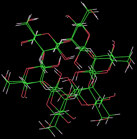
The elucidation of the 3D structure of native curdlan, has been under several investigations yielding somehow conflicting results. Previous investigations elicited the molecular structure as a six-fold
Fig. 3 Schematic representation of the structural packing observed in native curdlan I
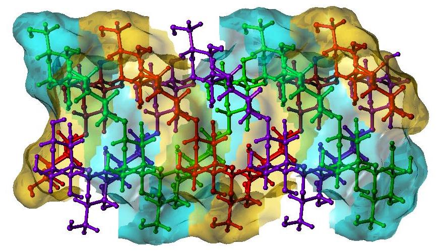
The hydrated form of Curdlan as analysed by fiber X-ray diffraction is organized as a triple helical structure of parallel right-handed strands (c value of 18.78 Å) packed in a triclinic unit cell
Fig.4 3D representation of the structure of the packing observed in curdlan II
The dehydrated form of curdlan is structurally well organized and gives rise to an X-ray pattern that can be analysed as an hexagonal unit cell, with space group P63
The packing of the chains in the unit cell is maintained by a series of strong hydrogen bonds (having distance between 2.70 to 2.75 Å).
|