![]()
A Database of Polysachharide 3D structures

| Home | |||||||
| About | |||||||
| User Guide | |||||||
| > Chitin & Chitosan Structures | |||||||
| Methods | |||||||
| References | |||||||
| Wiki | |||||||
| Contact us | |||||||
|
** External Links **
|
||||||
|
|||||||
Discover Polysaccharides |
Chitins & Chitosans
......................................................................................... Chitin is a polysaccharide composed of 1→4 linked 2-acetamido-2-deoxy-β-D-glucopyranose which plays the role of structural element of the outer skeleton of fungi, insects and crustaceans. It is rather narrowly distributed on the evolution map between cellulose and collagen. The yield of chitin in nature is thought to be second to that of cellulose. The identification of chitin was made by Braconnot in 1810; upon treating mushrooms Agaricus volvaceus with warm diluted alkali, resulted a production of ammonia through distillation with potassium hydroxide and the generation of acetic acid throughout its degradation with sulphuric acid
The occurrence and distribution of chitin resulted from many investigations, and in 1977 Muzzarelli reported its distribution (along with that of its derivative (chitosan) in the living species
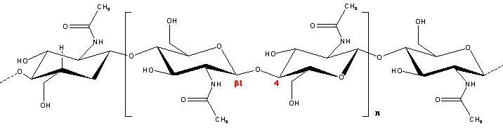
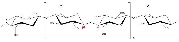
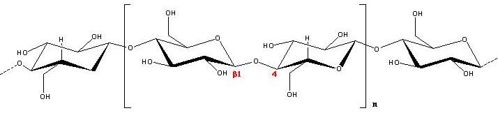
a. b. c. Fig. 1 Chemical structure of (a) Chitin, (b) Chitosan, and (c) Cellulose, the main polysaccharide components of supporting material in living things The biosynthesis route of chitin is well characterized in arthropods. In the de novo route, trehalose, one of the blood carbohydrate of insects is coverted in N-acetlyglucosamine-6-phosphate (GlcNAc-6-P) through several intermediate stages. GlcNAc-6-P is then converted into GlcNAc-uridine di-phosphate (UDP-GlcNAc) through GlcNAc-1-P. The UDP-GlcNAc is then transferred to form chitin poly-GlcNAc, by chitin synthetic enzyme. In this de novo route, trehalose is supplied freshly to synthesize chitin. There occurs another route, referred to as the salvage-route, whereby the sloughed cuticles are recycled. Consequently, little amino sugar is released into the environment as long as arthropods maintain an active metabolism. Native chitin occurs in fibrous crystalline states as several polymorphs. Three crystalline modifications, which are referred to as α, β and γ have been identified from X-ray diffraction studies. Of these, the α and β forms are analoguous to cellulose II and I, respectively, in terms of chain packing. Whereas their structural details have been characterized to varying extents, the γ form is still unresolved. α-chitin is the most abundant polymorph constituting various part of crustacean bodies such as outer skeletons, tendons and grasping pines of arrow worms. β chitin is the rarer form found in the squid pens, diatoms spines and tubes of pogonophora. β and γ chitins have been reported to transform into α-chitin by physical and chemical treatments. β-chitin exists as a crystalline hydrate of lower stability than the α-polymorph. γ-chitin is found in the cocoons of the beetles Ptinus tectus and Rhynchaenus fangi. In the family of β-chitin polymorphs the existence of one anhydrous form and two anhydrous forms has been reported
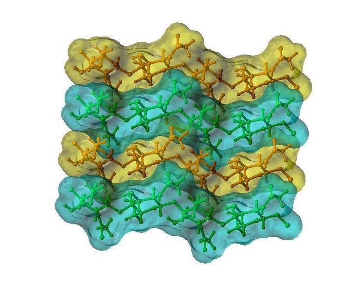
Fig. 2 3D representation of the crystal structure of β-chitin
Oriented fibers of highly crystalline, newer dried β-chitin have been obtained form tubes of Lamellibrachia satsuma and their structure characterized by synchrotron radiation X-ray fiber diffraction. Whereas the space group P21 remains unchanged, the unit cell dimensions change to a = 4.80 Å, b=11.15 Å, c (fiber repeat) = 10.44 Å and γ = 96.39 °. The increase in the volume of the unit cell indicates the presence of two water molecules. The crystal transition between the dihydrate and the anhydrous form was monitored by synchrotron radiation diffraction under controlled relative humidity. Such a transition can be reversible through an intermediate monohydrate form
The first model of the crystalline structure of the α-chitin was proposed by
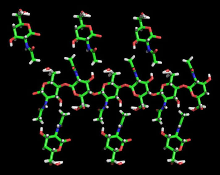
Fig. 3 3D representation of the crystal structure of α-chitin
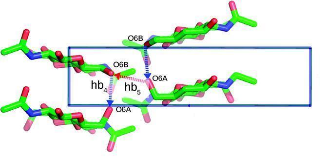
The hydroxymethyl groups at O-6 are statistically disordered, and the acetamido group is roughly perpendicular to the pyranose ring. Based on refined positions of the O-6 atoms, a network of hydrogen bonds involving these groups is found. It is proposed that the maximum number of hydrogen bonds will be formed in a situation where exist alternating O6 conformations in the chains adjacent along the a-axis. Such a network of hydrogen bonds can explain the main features of the polarized FTIR spectra of α-chitin.
Fig. 4 3D representation of the crystal structure of α-chitin (Courtesy: Pawel Sikorski; Ritsuko Hori; Masahisa Wada; Biomacromolecules 2009, 10, 1100-1105)
These chains are oriented in an anti-parallel fashion. Because of the polarity problem, the β-chitin to the α-chitin transition is also irreversible as in the case of cellulose. The natural occurrence of chitosan is limited to several organisms including Mycelia and sporangiophore of Phymoyces blakesleeanus, and the investigations of the crystalline structure have been very limited
|