![]()
A Database of Polysachharide 3D structures

| Home | |||||||
| About | |||||||
| User Guide | |||||||
| >α-D-Glucan Structures | |||||||
| Methods | |||||||
| References | |||||||
| Wiki | |||||||
| Contact us | |||||||
|
** External Links **
|
||||||
|
|||||||
Discover Polysaccharides |
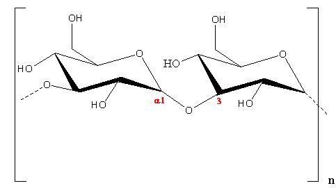
α-D-1,3-Glucan
......................................................................................... The polysaccharide α-D-1-3-glucan is mainly found in the cell wall of micro-organisms. Its occurrence was first detected in the cell walls of several non-pathogenic species and later on in and pathogenic ones (Aspergillus, Histoplasma, Cryptococcus neoformans etc.). In yeast, cell walls are critical for the maintenance of cell integrity, particularly in the face of challenges for its growth in mammalian hosts. Besides, it plays an important role in the virulence of multiple fungal pathogens. For example, such pathogenic fungus as Cryptococcus neoformans anchor their polysaccharide capsule to the cell surface via α-(1-3)-glucan in the wall. A linear form of the glucan is obtained from the cell walls of a number of fungi and yeasts where it is present in crystalline form. Some bacteria such as Streptococcus mutans and Streptococcus salivarius produce the branched glucan extracellularly in human saliva and a connection with the cause of dental carries has been suggested
Fig. 1 Schematic representation of the repeating unit of α-(1→3)-D-Glucan
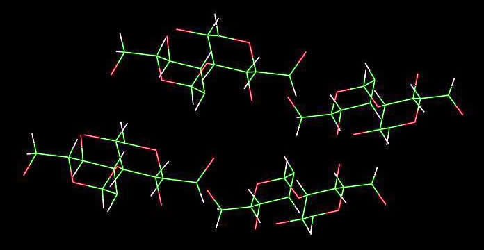
The orthorhombic unit cell having space group symmetry : P212121. Each helix occurs in a two-fold helical conformation, with a fiber repeat of 8.44 Å. This conformation is further stabilised by an intrachain hydrogen bond between O2-H and O-4 across the 1→3 glycosidic linkage. 4 chains cross the unit cell and they are arranged in an anti-parallel fashion
Fig. 2 Schematic representation of the 3D structure of α-(1→3)-D-Glucan They are arranged in a sheet-like fashion which resembles the packing features observed in cellulose II . |