![]()
A Database of Polysachharide 3D structures

| Home | |||||||
| About | |||||||
| User Guide | |||||||
| > Glucan Structures | |||||||
| Methods | |||||||
| References | |||||||
| Wiki | |||||||
| Contact us | |||||||
|
** External Links **
|
||||||
|
|||||||
Discover Polysaccharides |
α-D-1,3-Glucan triacetate
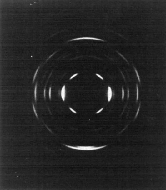
......................................................................................... The polysaccharide α-D-1→3-glucan is mainly found in the cell wall of micro-organisms. Its occurrence was first detected in the cell walls of several non-pathogenic species and later on in and pathogenic ones (Aspergillus, Histoplasma, Cryptococcus neoformans etc.). In yeast, cell walls are critical for the maintenance of cell integrity, particularly in the face of challenges for its growth in mammalian hosts. Besides, it plays an important role in the virulence of multiple fungal pathogens. For example, such pathogenic fungus as Cryptococcus neoformans anchor their polysaccharide capsule to the cell surface via α-(1→3)-glucan in the wall. A linear form of the glucan is obtained from the cell walls of a number of fungi and yeasts where it is present in crystalline form. Some bacteria such as Streptococcus mutans and Streptococcus salivarius produce the branched glucan extracellularly in human saliva and a connection with the cause of dental carries has been suggested As with other polysaccharides, there exist several crystalline polymorph of α-1→3 glucan. And the so-called regenerated form can be obtained through solid-state deacetylation of their acetyl derivative. After extraction from the fruiting body of a fungus, Laetiporus sulphureus, a pure α-1→3 glucan underwent complete acetylation with trifluoroacetic anhydride.
Fig. 1 The X-ray fibre pattern of α-(1→3)-D-Glucan triacetate. The fibre axis is vertical.
A susbsequent dissolution in chloroform yielded a film which was stretched in glycerin at 160°-180°C. A well-oriented film was annealed under tension in a sealed bomb in the presence of water and gave rise to an X-ray fiber diffraction pattern of sufficient quality. All the 41 observed X-ray reflections can be indexed on a monoclinic unit cell having dimensions of a=30.17, b= 17.42 and c = 12.11 Å (fiber axis) and β = 90°. The probable space group of P21 symmetry was assigned and a three dimensional model was established using the X-ray intensity combined with a stereochemical model refinement. The structural model is indicated by a residual index R= 26%. Six polysaccharide chains pass through the unit cell with alternating polarity. The chains are located in an hexagonal packing arrangement. The chain backbone conformation is a left-handed, three-fold helix, but all nine primary acetoxy groups are in non-equivalent rotational positions
|