Alginic acid
.........................................................................................
Introduction
Alginic acid, also called algin or alginate, is an anionic polysaccharide distributed widely in the cell walls of brown algae, where it, through binding water, forms a viscous gum. In extracted form it absorbs water quickly; it is capable of absorbing 200-300 times its own weight in water. Its colour ranges from white to yellowish-brown. It is sold in filamentous, granular or powdered forms.
Alginate can be produced by a microbial fermentation using bacteria such as Azobacter Vinelandii and Pseudomonas Aeruginosa
 .
These bacteria produce a polysaccharide with a structure resembling alginate, differing only in that there are acetyl groups on a portion of the C2 and C3 hydroxyls. It is believed that the acetate groups are associated mainly with the D-mannuronic acid residues
. The level of acetylation is variable as is the mannuronic and guluronic acid content.
.
These bacteria produce a polysaccharide with a structure resembling alginate, differing only in that there are acetyl groups on a portion of the C2 and C3 hydroxyls. It is believed that the acetate groups are associated mainly with the D-mannuronic acid residues
. The level of acetylation is variable as is the mannuronic and guluronic acid content.
The quick absorption of water makes alginate useful as an additive in dehydrated products such as slimming aids, and in the manufacture of paper and textiles. It is also used for waterproofing and fireproofing fabrics, as a gelling agent, and for thickening drinks, ice cream and cosmetics. Alginate is also used in various pharmaceutical preparations. Alginate is used extensively as an impression-making material in dentistry, prosthetics, lifecasting and occasionally for creating positives for small-scale casting. It is also used in the food industry, for thickening soups and jellies. Calcium alginate is used in different types of medical products, including burn dressings that promote healing and can be removed with less pain than conventional dressings. Also, due to alginate's biocompatibility and simple gelation with divalent cations such as Ca2+, it is widely used for cell immobilization and encapsulation. Alginic acid is also used in culinary arts, where natural juices of fruits and vegetables are encapsulated in bubbles that "explode" on the tongue when consumed. One of the most famous examples of this use of alginic acid was when Ferran Adrià used alginic acid to make apple caviar. Due to its ability to absorb water quickly, alginate can be changed through a lyophilization process to a new structure that has the ability to expand. It is used in the weight loss industry as an appetite suppressant. In March, 2010 researchers at Newcastle University announced that dietary alginates can reduce human fat uptake by more than 75%.
The composite structure of alginate is made up of blocks of beta-d-mannuronic acids (ManA) and blocks of α-l-guluronic acids (GulA). The composition and block lengths are species dependent. For example the sample from Fucus or Asophyllum species is 97% mannunoric acid, and that from Lamineria hyperborean is rich in polyguluronic acid. The presence of thes different types of polymer segment has been shown by mild acid hydrolysis
.
One type of segment consists entirely of guluronic acid residues, one consists entirely of mannuronic acid residues and the third consists of an alternating sequence of mannuronic and guluronic acid residues. Because the mild acid hydrolysis is not very selective it is difficult to say how long the segments are, but it appears that the average segment could be up to 80 residues long. Because GluA and ManA adopt 4C1 and 1C4 chair conformations, respectively, the morphology of the two constituting polysaccharides are quite unrelated.
(a) 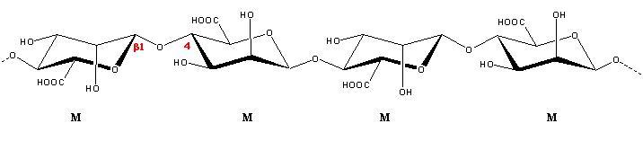
(b) 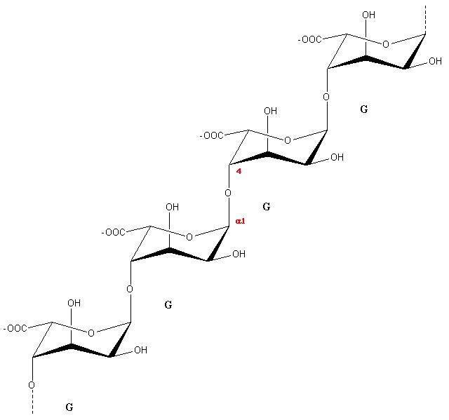
Fig. 1 Schematic representation of the repeating units found in
(a) β-D-mannuronic acids (ManA) and (b) blocks of α-L-guluronic acids (GulA).
Poly(β-D-Mannuronic acid)
The X-ray diffraction as recorded from a bundle of fibers prepared from Fucus vesiculosius are reminiscent of those from Mannan I
.
The elucidation of the structure was attempted using polarized infrared spectra. The molecule is a ribbon-like 2-fold helix exhibiting features characteristic of cellulose and mannan, with the occurrence of the OH3…O5 intrachain hydrogen bond (2.7 Å). Within the orthorhombic unit cell, the helices are arranged in an antiparallel fashion, with the occurrence of an intrachain hydrogen bond (O2H…O5) having a distance of 3.0 Å

Fig. 2 Three dimensional structure of Poly (β-D-Mannuronic acid).
Poly (α-L-Guluronic acid)
The structure of Poly (α-L-Guluronic acid) was also derived from a joint analysis
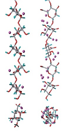
of X-ray diffraction data and polarized infra-red spectra. Within the X-ray diffraction pattern, the occurrence of the meridional reflection on the second layer line is diagnostic of a two-fold helix with a pitch of 8.7 Å, i.e. 1.7 Å shorter than in cellulose and mannan. This occurs from the fact that atoms O-1 and O-4 are both axial in the favored 1C4 chair conformation for guluronate residue. As a result, the molecular structure is more buckled than the familiar ribbon-like cellulose chain. Within the unit cell the polysaccharide chains are antiparallel. The pairwise association of helices mediated by an array of calcium ions in the middle bears certain resemblance to a Styrofoam box packed with eggs. This similarity has led to the so-called egg-box model
to depict the junction zone implicated in the alginate gelation process.
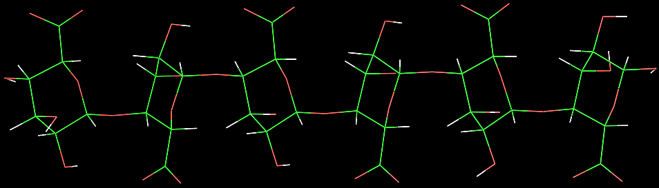
Fig. 3 Three dimensional structure of Poly (α-L-Guluronic acid).
Other fiber diffraction patterns have been obtained from different salts
.
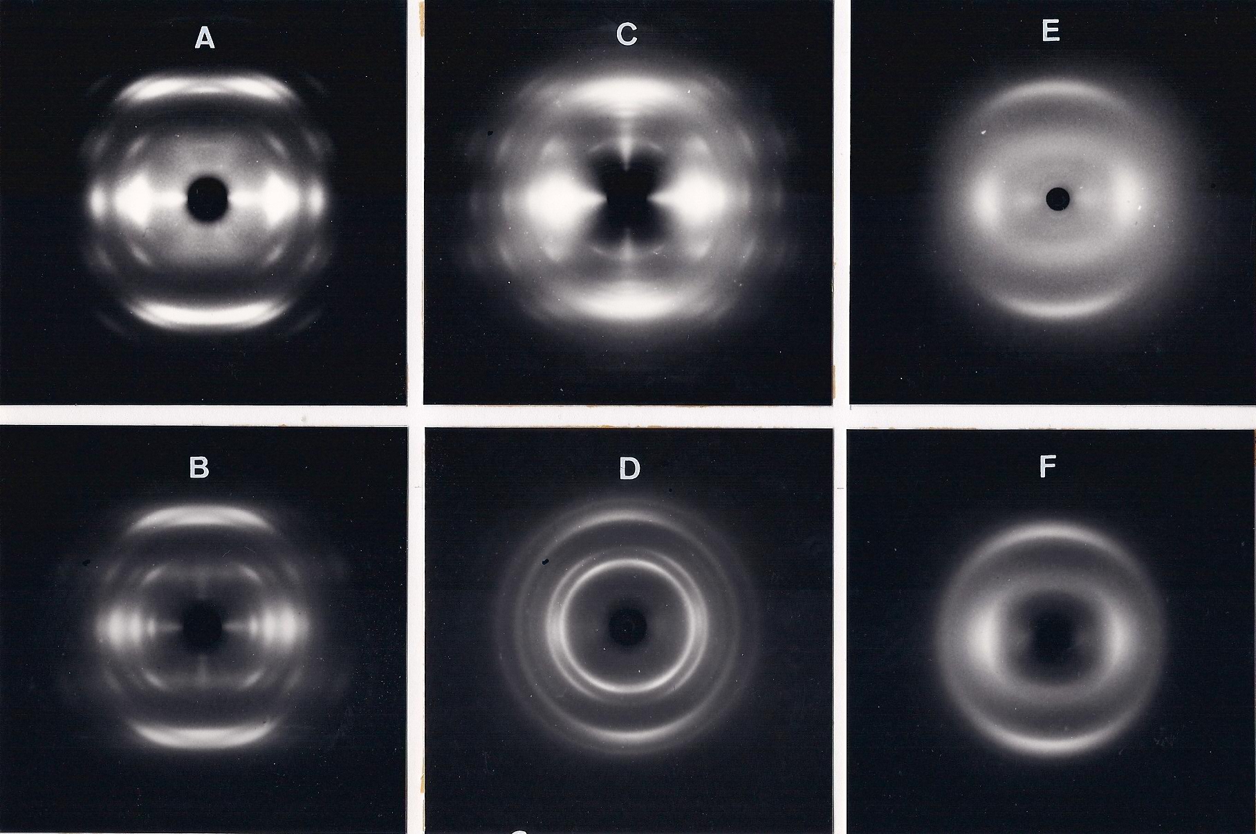
Fig. 4 X-ray fiber diffraction patterns obtained from Poly (α-L-Guluronic acid) and several salts. (A) polyguluronic acid, (B) polyguluronate-K+, (C) polyguluronate-NH4+, (D) polyguluronate-Li+, (E) polyguluronate-Ca2+, (F) polyguluronate-Mg2+.
With the exception of the calcium and the magnesium salts it is possible to measure layer line spacings of about 8.7 Å and a strong axial reflections of about 4.35 Å Thus it is apparent that the accepted two-fold helical chain conformation, having two sugars residues in the 1C4 chair conformation in the repeat is maintained in various salt forms in the condensed phase. The X-ray patterns shown are those obtained after annealing the specimens, when an increase in crystallinity is often observed. Before annealing, the X-ray patterns of a wide range of salt forms invariably have the same general appearance and distribution of intensity as shown by the Ca2+ and Mg2+ salts. It is therefore considered unlikely that the conformation of the calcium and magnesium polyguluronate deviate from the two fold helical conformation in the potassium form.The X-ray data indicates the occurrence of the two-fold symmetry of the chain (n=2) and average value of the advance per monomer of h=4.33 Å (range 4.30 to 4.40 Å).
The ionic polysaccharide alginate has a wide range of industrial applications that mainly rest on its ability to form gels in the presence of divalent cations such as calcium. In the case of alginate, gel formation has been demonstrated to result from a specific interaction between calcium ions and blocks of guluronate residues . Selectivity coefficients (KK Ca) indicate a strong calcium binding for polyguluronate chains compared to that of polymannuronate chains. These results demonstrate the importance of the structural features of the polysaccharide chains in calcium binding and indicate that differences between them result in some way from differences in the steric arrangement of the active groups. A structural study of all known single crystals of calcium carbohydrate complexes reported that calcium interactions display considerable stereospecificity and that the strongest binding is likely to result from sets of oxygen atoms that possess the proper spacing and geometry for substituting water in the calcium coordination shell
.
In this way, the axial–equatorial–axial sequence of hydroxyl groups on six-membered rings has been recognized as a very efficient pattern for calcium binding. Guluronic acid residues exhibit such a pattern. The calcium-chain interactions were screened for polyguluronic acic and polymannuronic acid in an attempt to understand the experimentally observed differences. For polysaccharide systems, gel formation is thought to result from chain association in which the chains assume an ordered secondary structure. In the case of alginates the cooperative nature of the calcium binding is best explained by the presence of the binding sites distributed in ordered arrays and formed between chains. The ordered conformations of the polysaccharides were used to study the calcium–oligouronate interaction. Since the calcium salts of polyguluronate and polymannuronate are known to exhibit, respectively, a 21 and a 32 helical conformation in the solid state, only these forms were explored. (Ref). Starting from the stable helical conformations of the polysaccharides, a calcium ion probe was moved around the van der Waals surface of the polymers. The interaction energies are significantly lower for the (α 1→4)-linked polymers than for the (β 1→4) ones. This indicates strong calcium binding for polyguluronate . This is in agreement with the experimental data. In order to evaluate the specificity of the interaction, the number of favorable positions for the calcium ion along the chain has been computed over a 15kcalmol -1 window above the lowest interaction energy. A representation of the polymer chains with these favorable binding sites is shown.
As previously suggested, the corrugated polyguluronate chains offer a proximity of several oxygen atoms from consecutive residues whose stereochemical arrangement fit into the coordination sphere of the cation. In contrast, the linear conformations of polymannuronate chains do not offer such a suitable arrangement. Consequently, the polyguluronate chains exhibit a high specificity for calcium binding and present well-defined chelation sites. The best interaction positions over the 15 kcal mol-1 window correspond to periodic sites (perfectly defined in the case of the guluronate chain with five periodic sites of the hexamer) which refer to an identical chelation site. It is interesting to note that the 31 conformation of the galacturonate chain provides a second possible binding site. In all cases, the (α 1→4)-linked polymers provide tetradentate chelation sites. As for the polygalacturonate chain, the coordination patterns predicted for the two helical conformations are in agreement with those derived from extended X-ray absorption fine structure (EXAFS) experiments for single pectate chains. Nevertheless, as calcium ion dimerizes the pectate and alginate chains in solution yielding gel formation, the four oxygen atoms predicted as ligands are not all expected to be involved in the coordination sphere of calcium ions at the dimer level.
The highly specific interaction described above clearly indicates that for (α 1→4)-linked polyuronates, calcium binding is far from a pure electrostatic interaction between cations and carboxylate oxygen atoms. In contrast, (β 1→4)-linked polymers seem to bind calcium essentially through electrostatic interactions. As shown, many positions for Ca2+ are equally favourable, the only necessary condition being the proximity of the negatively charged oxygen atoms. Consequently, polymannuronate do not display any specificity for calcium binding. The flat chains do not offer sites for preferential interactions to occur. This lack of specificity in calcium binding corroborates the absence of selectivity experimentally observed with respect to sodium ions.
α-L-(1→4)-GulA β-D-(1→4)-ManpA
Fig. 5 Schematic representation of the repeating unit of Guluronic acid & Mannuronic acid
A molecular modeling procedure has been developed which involves a pairing procedure that evaluates all the possible associations of the ordered polyuronate chains with calcium ions to form dimers. Starting from the stable ordered forms of polygalacturonate all possible ways to form Ca2+-bridged dimers are evaluated; the parallel and anti-parallel relative arrangements of the chains being also considered. The results indicate that, irrespective of the helical form, the antiparallel arrangement was the most favorable one. It allowed for an efficient connection of the chains, as illustrated by the shorter interhelical distance, and thus optimized the van der Waals contacts.
.
The results reported indicate that both parallel and anti-parallel arrangements of the chains provide efficient associations of polyguluronate chains. The specific interactions between the chains may be very different as illustrated by the best structures of each type shown in Fig. 4, parts (A) and (B). These represent the two general kinds of favorable association of 21 helical chains, and are symbolized “|” and “\” (by reference to the shape of the structures as viewed from the top). The energetic contributions were different in these two cases. The parallel chain pairing provided a good electrostatic contribution with calcium ions coordinated by five uronate oxygen atoms at distances ranging from 2.24 to 2.51 Å (Fig. 4a). The antiparallel chain pairing gave rise to an efficient hydrogen bond network with two complementary hydrogen bonds: O2…O6, 2.73 Å (2.88 Å), and O3…O5, 2.87 Å (2.91 Å). It also provided a unique periodic chelation site for calcium ions. It is not possible to discriminate between the two forms of these chain pairings based on total energy; however, the antiparallel arrangement is probably favored in the gel since it has been reported that urea substantially weakens the gel indicating a notable contribution of hydrogen bonds to gel strength. Moreover, the antiparallel association of 21 helical chains is the arrangement found in the solid state. The shape of this structure is close to that of the popular “egg box model”. This model, as commonly represented, was evaluated by searching for the best way to connect two anti-parallel chains having the glycosidic oxygen atoms face to face (no shift of one chain with respect to the other). The results are given in Table 1 and the van der Waals representation of the corresponding structure is given in Fig. 6a. Both energetic and structural considerations indicate that the polyguluronate “egg box” structure corresponds to a favorable association. It provides a compact dimer, with cavities well adapted to calcium ions (Fig. 6a), resulting in an efficient junction zone. Nevertheless, the model is wrong in the description of calcium coordination: only four ligands from the chains, O2 and O6 of both chains and not 10 oxygen atoms, bind the calcium ion. The other postulated ligands, O3, O4, and O5, are at distances longer than 3 Å. The involvement of these atoms in the coordination sphere of calcium ions would lead to an inter-penetration of the two chains. In fact, as described by Smidrød and Draget,21 “the egg box model may be regarded as principally correct, giving an intuitive understanding of the characteristic chelate-type of the ion-binding properties of alginates”.
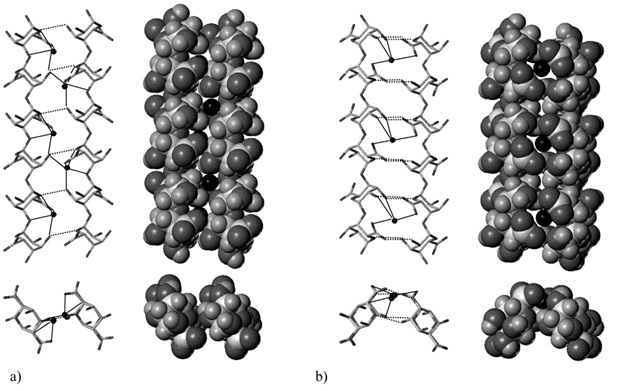
Fig. 6 Representations (stick and van der Waals structures) of the best [Ca2+ chain] associations of 2-fold guluronate chains: (a) parallel arrangement; (b) antiparallel arrangement. Positions of calcium ions have been reoptimized with respect to the dimer structures. Dark
circles represent calcium ions. Key: (s) calcium coordination; (- - -) hydrogen bonds.