![]()
A Database of Polysachharide 3D structures
| Home | |||||||
| About | |||||||
| User Guide | |||||||
| > Hyaluronate II Potassium for EXPERTS | |||||||
| Methods | |||||||
| References | |||||||
| Wiki | |||||||
| Contact us | |||||||
|
|
|||||||
Polysaccharides For Experts |
Hyaluronate II Potassium.........................................................................................
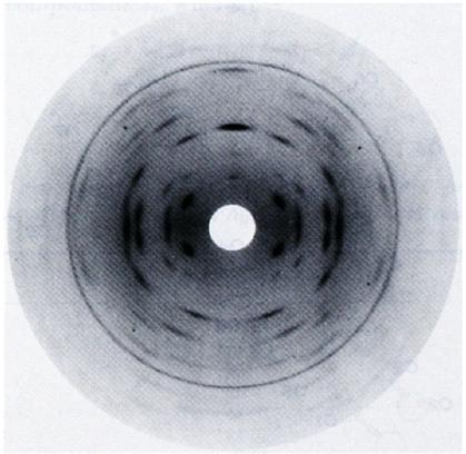
Fig.1 The diffraction pattern of potassium hyaluronate. Meridional reflections are seen on every 4th layer-line indicating a 4-fold helix. *Permission Pending for Diffraction Diagrams
......................................................................................... Hyaluronate II Potassium Space Group : tetragonal P43212 Unit Cell Dimensions (a, b, c in Å and α, β, γ in °) (No values are displayed if no information is available or the value is zero) a (Å) = 9.96 - b (Å) = 9.96 - c (Å) = 37.88 Helix Type : two anti-parallel, extended 4-fold helical chains Method(s) Used For Structure Determination : X-ray diffraction Link to the Abstract : http://www.ncbi.nlm.nih.gov/pubmed/6631953
Download Structure
Unit hyaluronic-acid-2-K_expanded
|