![]()
A Database of Polysachharide 3D structures
| Home | |||||||
| About | |||||||
| User Guide | |||||||
| > Hyaluronate III Sodium for EXPERTS | |||||||
| Methods | |||||||
| References | |||||||
| Wiki | |||||||
| Contact us | |||||||
|
|
|||||||
Polysaccharides For Experts |
Hyaluronate III Sodium.........................................................................................
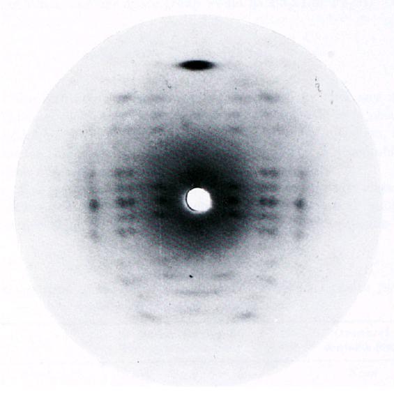
Fig.1 Diffraction pattern from a three-fold sodium hyaluronate. The unit cell dimensions are a=b=11.70, c (fiber axis)= 28.50 and γ=120°. The meridional direction is vertical and the sample was tilted from a direction normal to the beam by 15°. *Permission Pending for Diffraction Diagrams
......................................................................................... Hyaluronate III Sodium Unit Cell Dimensions (a, b, c in Å and α, β, γ in °) (No values are displayed if no information is available or the value is zero) a (Å) = 11.70 - b (Å) = 11.70 - c (Å) = 28.50 - γ (°) = 120.00 Helix Type : regular, left-handed 3-fold helix Link to the Abstract : http://www.ncbi.nlm.nih.gov/pubmed/1206703
Download Structure
|