![]()
A Database of Polysachharide 3D structures
| Home | |||||||
| About | |||||||
| User Guide | |||||||
| > Dermatan 4 sulphate II Na (Sodium dermatan 4-sulphate II) (allomorph II) for EXPERTS | |||||||
| Methods | |||||||
| References | |||||||
| Wiki | |||||||
| Contact us | |||||||
|
|
|||||||
Polysaccharides For Experts |
Dermatan 4 sulphate II Na (Sodium dermatan 4-sulphate II) (allomorph II).........................................................................................
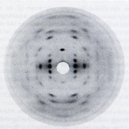
Fig.1 Fiber diffraction patterns of sodium dermatan sulfate (a)Trigonal form with meridional reflexions on l = 3n. *Permission Pending for Diffraction Diagrams
......................................................................................... Dermatan 4 sulphate II Na (Sodium dermatan 4-sulphate II) (allomorph II) Space Group : allomorph II : trigonal P3221 Unit Cell Dimensions (a, b, c in Å and α, β, γ in °) (No values are displayed if no information is available or the value is zero) a (Å) = 14.60 - b (Å) = 14.60 - c (Å) = 28.23 Method(s) Used For Structure Determination : X-ray diffraction Link to the Abstract : http://www.ncbi.nlm.nih.gov/pubmed/6631956
Download Structure
Unit dermatan-32-allo2_expanded
|