![]()
A Database of Polysachharide 3D structures
| Home | |||||||
| About | |||||||
| User Guide | |||||||
| > Poly-β-D-Mannuronic Acid (Alginic acid) for EXPERTS | |||||||
| Methods | |||||||
| References | |||||||
| Wiki | |||||||
| Contact us | |||||||
|
|
|||||||
Polysaccharides For Experts |
Poly-β-D-Mannuronic Acid (Alginic acid).........................................................................................
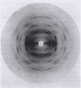
Fig.1 X-ray fiber diffraction photograph obtained from polymannuronic acid. The pattern indexes on an orthorhombic unit cell with a layer line spacing of 10.4 Å. The fiber axis is vertical. *Permission Pending for Diffraction Diagrams
......................................................................................... Poly-β-D-Mannuronic Acid (Alginic acid) Space Group : orthorhombic P212121 Unit Cell Dimensions (a, b, c in Å and α, β, γ in °) (No values are displayed if no information is available or the value is zero) a (Å) = 7.60 - b (Å) = 10.40 - c (Å) = 8.60 Method(s) Used For Structure Determination : X-ray diffraction and polarized infrared studies Link to the Abstract : http://www.ncbi.nlm.nih.gov/pubmed/4733712 Structural Components of Alginic Acid. I. The Crystalline Structure of Poly-?-D-Mannuronic Acid. Results of X-Ray Diffraction and Polarized Infrared StudiesAtkins, E.D.T., Nieduszynski, I.A., Mackie, W., Parker, K.D. and Smolko, E.E., Biopolymers, 1973, 12(8), Pages 1865 - 1878
Download Structure
Unit mannuronic-acid-32-Ca_expanded
|