![]()
A Database of Polysachharide 3D structures
| Home | |||||||
| About | |||||||
| User Guide | |||||||
| > Chitin II (chitin α) for EXPERTS | |||||||
| Methods | |||||||
| References | |||||||
| Wiki | |||||||
| Contact us | |||||||
|
|
|||||||
Polysaccharides For Experts |
Chitin II (chitin α).........................................................................................
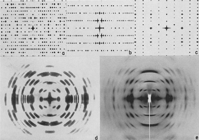
Fig.1 Optically derived Fourier Transforms (diffractograms) from the (100), (010) and (001) projections (a), (b) and (c) respectively, of the best model of chitin. (d) is the composite diffractogram of (a), (b) and (c) with the reflections of (b) transferred to the equator; (e) the disorientation and the fading off have been introduced artificially in order to match the X-ray diffraction pattern. The fiber axis is vertical except in (b) where it is perpendicular to the plane of the figure. *Permission Pending for Diffraction Diagrams
......................................................................................... Chitin II (chitin α) Unit Cell Dimensions (a, b, c in Å and α, β, γ in °) (No values are displayed if no information is available or the value is zero) a (Å) = 4.74 - b (Å) = 18.86 - c (Å) = 10.32 - γ (°) = 90.00 Method(s) Used For Structure Determination : X-ray diffraction Link to the Abstract : http://www.ncbi.nlm.nih.gov/pmc/articles/PMC2224123/
Download Structure
|