A Database of Polysachharide 3D structures

| Home | |||||||
| About | |||||||
| User Guide | |||||||
| > Cellulose IV1 for EXPERTS | |||||||
| Methods | |||||||
| References | |||||||
| Wiki | |||||||
| Contact us | |||||||
|
|
|||||||
Polysaccharides For Experts |
Cellulose IV1.........................................................................................
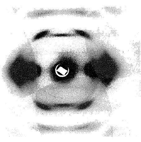
Fig.1 X-ray diffraction diagrams of cellulose IV1. Fibre axis is vertical. *Permission Pending for Diffraction Diagrams
......................................................................................... Cellulose IV1 Unit Cell Dimensions (a, b, c in Å and α, β, γ in °) (No values are displayed if no information is available or the value is zero) a (Å) = 8.03 - b (Å) = 8.13 - c (Å) = 10.34 - γ (°) = 90.00 - β (°) = 90.00 - α (°) = 90.00 Method(s) Used For Structure Determination : X-ray diffraction and stereochemical model refinement Link to the Abstract : http://rparticle.web-p.cisti.nrc.ca/rparticle/AbstractTemplateServlet?calyLang=eng&journal=cjc&volume=63&year=1985&issue=1&msno=v85-027
Download Structure
Unit cellulose-IV-1-cornerchain_unit cellulose-IV-1-2chains_expanded
|
