![]()
A Database of Polysachharide 3D structures
| Home | |||||||
| About | |||||||
| User Guide | |||||||
| > Cellulose III1 for EXPERTS | |||||||
| Methods | |||||||
| References | |||||||
| Wiki | |||||||
| Contact us | |||||||
|
|
|||||||
Polysaccharides For Experts |
Cellulose III1.........................................................................................
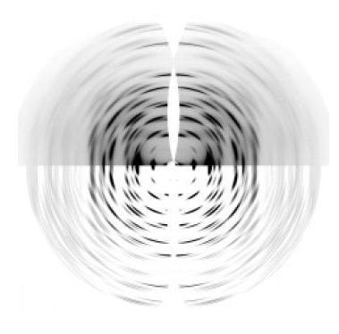
Fig.1 TOP : synchrotron X-ray diffraction data collected on an on-line MAR image plate from an oriented sample of cellulose III1 on station ID2A at the ESRF, Grenoble France. BOTTOM : a fit of the top pattern obtained during the measurement of the Bragg intensities and background using software that takes into account fiber texture. Comparison of the top and bottom sections indicates the agreement between observed and measured Bragg intensities. *Permission Pending for Diffraction Diagrams
Fig.1 TOP : synchrotron X-ray diffraction data collected on an on-line MAR image plate from an oriented sample of cellulose III1 on station ID2A at the ESRF, Grenoble France. BOTTOM : a fit of the top pattern obtained during the measurement of the Bragg intensities and background using software that takes into account fiber texture. Comparison of the top and bottom sections indicates the agreement between observed and measured Bragg intensities.
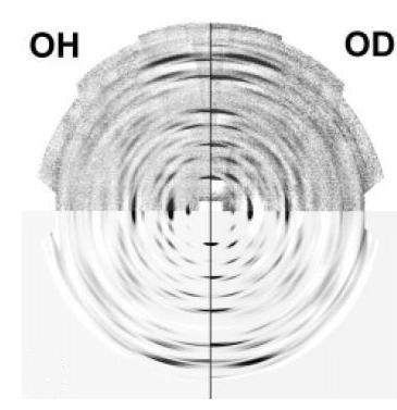
Fig.2 TOP : neutron fiber diffraction patterns collected from oriented samples of cellulose III1, one hydrogenated (LEFT-HAND QUADRANT) and the other deuterated (RIGHT-HAND QUADRANT). The BOTTOM QUADRANTS show 3D fits of the Bragg intensities using software that takes into account fiber texture. The images in the Cellulose III1 have been re-mapped into cylindrical reciprocal space with the fiber axis vertical. *Permission Pending for Diffraction Diagrams
......................................................................................... Cellulose III1 Unit Cell Dimensions (a, b, c in Å and α, β, γ in °) (No values are displayed if no information is available or the value is zero) a (Å) = 4.45 - b (Å) = 7.85 - c (Å) = 10.31 - γ (°) = 105.10 - β (°) = 90.00 - α (°) = 90.00 Helix Type : parallel arrangement, one-chain Method(s) Used For Structure Determination : X-ray and neutron fiber diffraction Link to the Abstract : http://pubs.acs.org/doi/abs/10.1021/ma0485585
Download Structure
|