![]()
A Database of Polysachharide 3D structures
| Home | |||||||
| About | |||||||
| User Guide | |||||||
| > Cellulose 1β for EXPERTS | |||||||
| Methods | |||||||
| References | |||||||
| Wiki | |||||||
| Contact us | |||||||
|
|
|||||||
Polysaccharides For Experts |
Cellulose 1β.........................................................................................
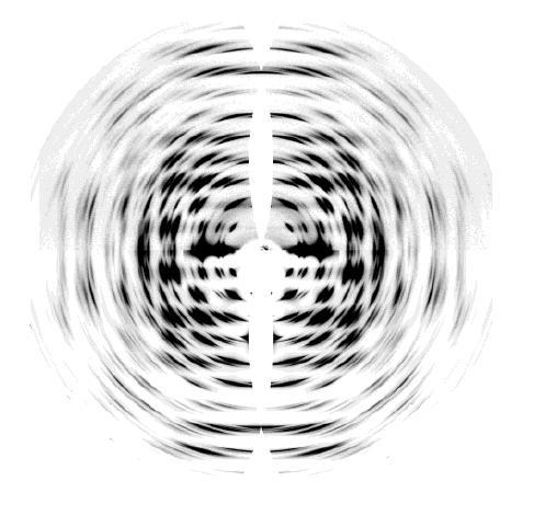
Fig.1 (TOP) Synchrotron X-ray diffraction data collected on an online MAR image plate from fibers of Halocynthia cellulose Iα on station ID2A at the ESRF, Grenoble, France. (BOTTOM) A 3D fit of the Bragg intensities, done using custom-written software that takes into account fiber texture. The images have been remapped into cylindrical reciprocal space with the fiber axis vertical. *Permission Pending for Diffraction Diagrams
Fig.1 (TOP) Synchrotron X-ray diffraction data collected on an online MAR image plate from fibers of Halocynthia cellulose Iα on station ID2A at the ESRF, Grenoble, France. (BOTTOM) A 3D fit of the Bragg intensities, done using custom-written software that takes into account fiber texture. The images have been remapped into cylindrical reciprocal space with the fiber axis vertical.
Fig.2 Neutron fiber diffraction patterns collected from two fibers of Halocynthia cellulose Iβ , one hydrogenated (TOP LEFT-HAND QUADRANT) and the other deuterated (TOP RIGHT-HAND QUADRANT). The bottom quadrants show 3D fits of the Bragg intensities, done using custom-written software that takes into account fiber texture. The images have been remapped into cylindrical reciprocal space with the fiber axis vertical. *Permission Pending for Diffraction Diagrams
......................................................................................... Cellulose 1β Unit Cell Dimensions (a, b, c in Å and α, β, γ in °) (No values are displayed if no information is available or the value is zero) a (Å) = 7.78 - b (Å) = 8.20 - c (Å) = 10.38 - γ (°) = 96.55 - β (°) = 90.00 - α (°) = 90.00 Helix Type : parallel-up chain arrangement, two parallel chains Method(s) Used For Structure Determination : X-ray and neutron fiber diffraction Link to the Abstract : http://www.ncbi.nlm.nih.gov/pubmed/12149011
Download Structure
Unit cellulose-1-beta-cornerchain_unit cellulose-1-beta-2chains_expanded
|