![]()
A Database of Polysachharide 3D structures
| Home | |||||||
| About | |||||||
| User Guide | |||||||
| > Carrageenan Iota for EXPERTS | |||||||
| Methods | |||||||
| References | |||||||
| Wiki | |||||||
| Contact us | |||||||
|
|
|||||||
Polysaccharides For Experts |
Carrageenan Iota.........................................................................................
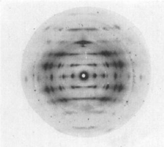
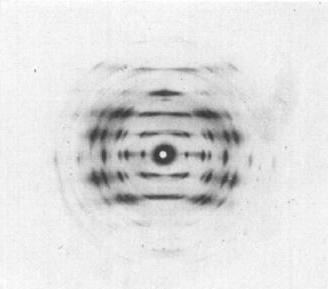
Fig.1 The fibres were tilted out of the plane normal to the X-ray beam 10° in order to record the 003 reflecrion (0.443 nm spacing). The specimen-to-film distance was approx. 30 mm. Exposure times were 23 h for the (Ca2+salt and 11 h for the Sr2 + salt). The relative humidky was maintained at 75%. Fibre diffraction patterns of the : (a)Fibre diffraction patterns of the Ca2+ i-carrageenate. *Permission Pending for Diffraction Diagrams
Fig.1 The fibres were tilted out of the plane normal to the X-ray beam 10° in order to record the 003 reflecrion (0.443 nm spacing). The specimen-to-film distance was approx. 30 mm. Exposure times were 23 h for the (Ca2+salt and 11 h for the Sr2 + salt). The relative humidky was maintained at 75%. Fibre diffraction patterns of the : (a)Fibre diffraction patterns of the Ca2+ i-carrageenate. (b)Fibre diffraction patterns of the Sr2+ i-carrageenate. *Permission Pending for Diffraction Diagrams
Fig.1 The fibres were tilted out of the plane normal to the X-ray beam 10° in order to record the 003 reflecrion (0.443 nm spacing). The specimen-to-film distance was approx. 30 mm. Exposure times were 23 h for the (Ca2+salt and 11 h for the Sr2 + salt). The relative humidky was maintained at 75%. Fibre diffraction patterns of the : (a)Fibre diffraction patterns of the Ca2+ i-carrageenate. (b)Fibre diffraction patterns of the Sr2+ i-carrageenate.
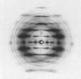
Fig.2 Fibre diffraction patterns of the Mg2+ i-carrageenate: (a)The normal type obtained from a fibre prepared under slow drying conditions. *Permission Pending for Diffraction Diagrams
Fig.1 The fibres were tilted out of the plane normal to the X-ray beam 10° in order to record the 003 reflecrion (0.443 nm spacing). The specimen-to-film distance was approx. 30 mm. Exposure times were 23 h for the (Ca2+salt and 11 h for the Sr2 + salt). The relative humidky was maintained at 75%. Fibre diffraction patterns of the : (a)Fibre diffraction patterns of the Ca2+ i-carrageenate. (b)Fibre diffraction patterns of the Sr2+ i-carrageenate.
Fig.2 Fibre diffraction patterns of the Mg2+ i-carrageenate: (a)The normal type obtained from a fibre prepared under slow drying conditions. (b)The unusual type with interleaving Iayer lines obtained from fibres prepared under rapid drying conditions. *Permission Pending for Diffraction Diagrams
......................................................................................... Carrageenan Iota Unit Cell Dimensions (a, b, c in Å and α, β, γ in °) (No values are displayed if no information is available or the value is zero) a (Å) = 13.73 - b (Å) = 13.73 - c (Å) = 13.28 - γ (°) = 120.00 Helix Type : two, identical, right. handed, 3-fold helical polysaccharide chains Method(s) Used For Structure Determination : X-ray diffraction Link to the Abstract : http://www.ncbi.nlm.nih.gov/pubmed/4453016
Download Structure
Unit carrageenan-iota_expanded
|