![]()
A Database of Polysachharide 3D structures
| Home | |||||||
| About | |||||||
| User Guide | |||||||
| > Xanthan for EXPERTS | |||||||
| Methods | |||||||
| References | |||||||
| Wiki | |||||||
| Contact us | |||||||
|
|
|||||||
Polysaccharides For Experts |
Xanthan.........................................................................................
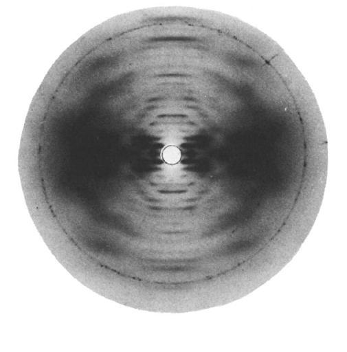
Fig.1 Diffraction pattern typical for both Xanthomonas campestrisand Xanthomonas phaseolipolysaccharides showing five-fold helical symmetry. The sharp Bragg reflection on the equator has a spacing of 1.9 nm. *Permission Pending for Diffraction Diagrams
......................................................................................... Xanthan Unit Cell Dimensions (a, b, c in Å and α, β, γ in °) (No values are displayed if no information is available or the value is zero) a (Å) = 29.00 - b (Å) = 24.90 - c (Å) = 47.00 - γ (°) = 90.00 Method(s) Used For Structure Determination : X-ray diffraction and computer-aided model building Link to the Abstract : http://pubs.acs.org/doi/abs/10.1021/bk-1977-0045.ch007 Xanthan Gum?Molecular Conformation and InteractionsMoorhouse, R., Walkinshaw, M.D. and Arnott, S., Acs. Symp. Ser. 1977, 45, 90-102
Download Structure
|