![]()
A Database of Polysachharide 3D structures
| Home | |||||||
| About | |||||||
| User Guide | |||||||
| > RMDP17 for EXPERTS | |||||||
| Methods | |||||||
| References | |||||||
| Wiki | |||||||
| Contact us | |||||||
|
|
|||||||
Polysaccharides For Experts |
RMDP17.........................................................................................
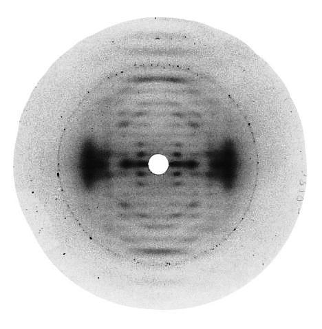
Fig.1 Diffraction pattern from a well oriented fiber of the sodium salt of RMDP17 kept at 75% relative humidity shows moderate polycrystallinity. The fiber is slightly tilted towards the incident X-ray beam (Cu Kα radiation, wavelength 1.5418 Å). The calcite ring corresponds to d-spacing 3.035 Å. *Permission Pending for Diffraction Diagrams
......................................................................................... RMDP17 Linkage : beta 1-3, beta 1-4 alpha 1-6 Unit Cell Dimensions (a, b, c in Å and α, β, γ in °) (No values are displayed if no information is available or the value is zero) a (Å) = 17.60 - c (Å) = 28.70 Helix Type : threefold helix symmetry Method(s) Used For Structure Determination : X-ray diffraction and computer-aided model building Link to the Abstract : Molecular structure of the rhamsan-like exocellular polysaccharide RMDP17 from Sphingomonas paucimobilis
Download Structure
|