![]()
A Database of Polysachharide 3D structures
| Home | |||||||
| About | |||||||
| User Guide | |||||||
| > Gellan Native K (Potassium Native Gellan) (Native Gellan-K ) for EXPERTS | |||||||
| Methods | |||||||
| References | |||||||
| Wiki | |||||||
| Contact us | |||||||
|
|
|||||||
Polysaccharides For Experts |
Gellan Native K (Potassium Native Gellan) (Native Gellan-K ).........................................................................................
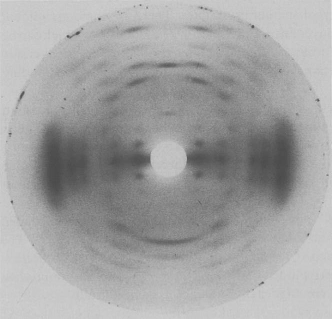
Fig.1 X-Ray diffraction pattern from a well oriented and polycrystalline fiber of the potassiumm salt of native gellan maintained at 75% relative humidity *Permission Pending for Diffraction Diagrams
......................................................................................... Gellan Native K (Potassium Native Gellan) (Native Gellan-K ) Unit Cell Dimensions (a, b, c in Å and α, β, γ in °) (No values are displayed if no information is available or the value is zero) a (Å) = 16.47 - b (Å) = 16.47 - c (Å) = 28.42 - γ (°) = 120.00 Helix Type : half-staggered, parallel, double helix Method(s) Used For Structure Determination : X-ray diffraction Link to the Abstract : http://www.ncbi.nlm.nih.gov/pubmed/1591755
Download Structure
|