![]()
A Database of Polysachharide 3D structures
| Home | |||||||
| About | |||||||
| User Guide | |||||||
| > M41 Capsular Polysaccharide (Escherichia coli ) for EXPERTS | |||||||
| Methods | |||||||
| References | |||||||
| Wiki | |||||||
| Contact us | |||||||
|
|
|||||||
Polysaccharides For Experts |
M41 Capsular Polysaccharide (Escherichia coli ).........................................................................................
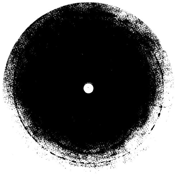
Fig.1 Diffraction pattern from 2-fold E. coli M41 polysaccharide. Two chains pass through the orthorhombic unit cell with dimensions a = 2.03 nm, b = 1.178 nm, and c (fiber axis) = 3.04 nm. The meridional direction is vertical and the sample was tilted from a direction normal to the beam by 9°. *Permission Pending for Diffraction Diagrams
......................................................................................... M41 Capsular Polysaccharide (Escherichia coli ) Linkage : alpha 1-2, beta 1-3, beta 1-4 Space Group : orthorhombic P212121 Unit Cell Dimensions (a, b, c in Å and α, β, γ in °) (No values are displayed if no information is available or the value is zero) Helix Type : antiparallel 2-fold helices Method(s) Used For Structure Determination : X-ray diffraction Link to the Abstract : http://www.ncbi.nlm.nih.gov/pubmed/319241
Download Structure
|