A Database of Polysachharide 3D structures

| Home | |||||||
| About | |||||||
| User Guide | |||||||
| > Dextran (high T polymorph) for EXPERTS | |||||||
| Methods | |||||||
| References | |||||||
| Wiki | |||||||
| Contact us | |||||||
|
|
|||||||
Polysaccharides For Experts |
Dextran (high T polymorph).........................................................................................
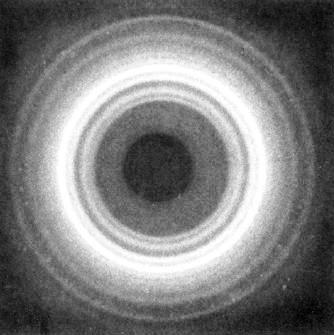
Fig.1 X-ray diffraction diagram of anhydrous dextran (flat film, under vacuum). *Permission Pending for Diffraction Diagrams
......................................................................................... Dextran (high T polymorph) Unit Cell Dimensions (a, b, c in Å and α, β, γ in °) (No values are displayed if no information is available or the value is zero) a (Å) = 9.22 - b (Å) = 9.22 - c (Å) = 7.78 - β (°) = 91.30 Helix Type : Two antiparallel-packed molecular chains Link to the Abstract : http://pubs.acs.org/doi/abs/10.1021/ma00131a017
Download Structure
Unit dextran-highT-anhyd_expanded
|
