![]()
A Database of Polysachharide 3D structures
| Home | |||||||
| About | |||||||
| User Guide | |||||||
| > Amylose V and propanol complex for EXPERTS | |||||||
| Methods | |||||||
| References | |||||||
| Wiki | |||||||
| Contact us | |||||||
|
|
|||||||
Polysaccharides For Experts |
Amylose V and propanol complex.........................................................................................
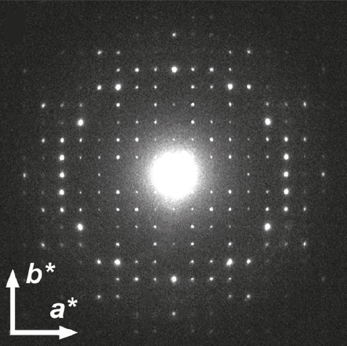
Fig.1 Electron diffraction diagram recorded on one square micrometer of the sample under frozen hydrated conditions, properly oriented with respect to the crystal. *Permission Pending for Diffraction Diagrams
......................................................................................... Amylose V and propanol complex Unit Cell Dimensions (a, b, c in Å and α, β, γ in °) (No values are displayed if no information is available or the value is zero) a (Å) = 28.26 - b (Å) = 29.50 - c (Å) = 8.01 - γ (°) = 90.00 Method(s) Used For Structure Determination : X-ray diffraction Link to the Abstract : http://pubs.acs.org/doi/abs/10.1021/ma101794w
Download Structure
Unit amylose-isopropanol_model2 amylose-isopropanol_model3
|