![]()
A Database of Polysachharide 3D structures
| Home | |||||||
| About | |||||||
| User Guide | |||||||
| > Amylose V for EXPERTS | |||||||
| Methods | |||||||
| References | |||||||
| Wiki | |||||||
| Contact us | |||||||
|
|
|||||||
Polysaccharides For Experts |
Amylose V.........................................................................................
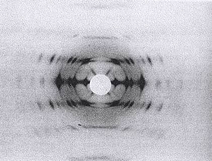
Fig.1 X-ray diffraction pattern of V a -amylose. *Permission Pending for Diffraction Diagrams
......................................................................................... Amylose V Unit Cell Dimensions (a, b, c in Å and α, β, γ in °) (No values are displayed if no information is available or the value is zero) a (Å) = 12.97 - b (Å) = 22.46 - c (Å) = 7.91 - γ (°) = 90.00 Helix Type : left-handed sixfold helix Method(s) Used For Structure Determination : X-ray diffraction Link to the Abstract : http://onlinelibrary.wiley.com/doi/10.1002/bip.1974.360130715/abstract Crystal and Molecular Structure of V-Anhydrous AmyloseWinter, W.T. and Sarko, A., 1974, Biopolymers, 13, 1447-1460
Download Structure
|