![]()
A Database of Polysachharide 3D structures
| Home | |||||||
| About | |||||||
| User Guide | |||||||
| > α 1→3-glucan for EXPERTS | |||||||
| Methods | |||||||
| References | |||||||
| Wiki | |||||||
| Contact us | |||||||
|
|
|||||||
Polysaccharides For Experts |
α 1→3-glucan.........................................................................................
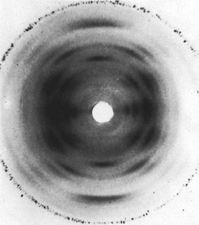
Fig.1 The fibre X-ray diffraction pattern of (I→3)-α-D-glucan. The fibre axis is vertical and the calibration line is the d= 2.319 Å line of NaF. *Permission Pending for Diffraction Diagrams
......................................................................................... α 1→3-glucan Space Group : orthorhombic P212121 Unit Cell Dimensions (a, b, c in Å and α, β, γ in °) (No values are displayed if no information is available or the value is zero) a (Å) = 16.46 - b (Å) = 9.55 - c (Å) = 8.44 Method(s) Used For Structure Determination : X-ray diffraction and stereochemical model refinement Link to the Abstract : http://www.sciencedirect.com/science/article/pii/0141813081900210
Download Structure
|