![]()
A Database of Polysachharide 3D structures
| Home | |||||||
| About | |||||||
| User Guide | |||||||
| > Nigeran for EXPERTS | |||||||
| Methods | |||||||
| References | |||||||
| Wiki | |||||||
| Contact us | |||||||
|
|
|||||||
Polysaccharides For Experts |
Nigeran.........................................................................................
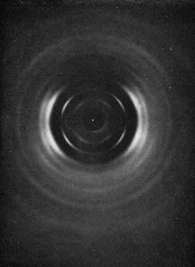
Fig.1 Fiber diffraction pattern of anhydrous nigeran. *Permission Pending for Diffraction Diagrams
Fig.1 Fiber diffraction pattern of anhydrous nigeran.
Fig.2 Electron diffraction pattern of single crystal of anhydrous nigeran. *Permission Pending for Diffraction Diagrams
......................................................................................... Nigeran Unit Cell Dimensions (a, b, c in Å and α, β, γ in °) (No values are displayed if no information is available or the value is zero) a (Å) = 17.76 - b (Å) = 6.00 - c (Å) = 14.62 - γ (°) = 90.00 Link to the Abstract : http://www.ncbi.nlm.nih.gov/pubmed/448733
Download Structure nigeran_xyz-expanded
|