![]()
A Database of Polysachharide 3D structures
| Home | |||||||
| About | |||||||
| User Guide | |||||||
| > Xylan (β 1→3) for EXPERTS | |||||||
| Methods | |||||||
| References | |||||||
| Wiki | |||||||
| Contact us | |||||||
|
|
|||||||
Polysaccharides For Experts |
Xylan (β 1→3).........................................................................................
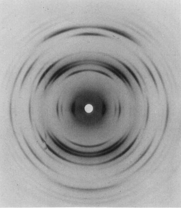
Fig.1 X-ray diffraction photograph obtained from the inner parts of the cell wall of Penicillus dumetosus. Cell axis vertical, X-ray beam almost normal to wall surfaces. The cell axis has been tilted to bring the 3.06 Å reflection on to the meridian. The specimen was maintained at 98% relative humidity during exposure. *Permission Pending for Diffraction Diagrams
......................................................................................... Xylan (β 1→3) Unit Cell Dimensions (a, b, c in Å and α, β, γ in °) (No values are displayed if no information is available or the value is zero) a (Å) = 15.40 - b (Å) = 15.40 - c (Å) = 6.12 - β (°) = 120.00 Helix Type : one right-handed triple helice per unit cell Method(s) Used For Structure Determination : X-ray diffraction Link to the Abstract : http://onlinelibrary.wiley.com/doi/10.1002/polc.5070280109/abstract
Download Structure
|