A Database of Polysachharide 3D structures

| Home | |||||||
| About | |||||||
| User Guide | |||||||
| > Scleroglucan for EXPERTS | |||||||
| Methods | |||||||
| References | |||||||
| Wiki | |||||||
| Contact us | |||||||
|
|
|||||||
Polysaccharides For Experts |
Scleroglucan.........................................................................................
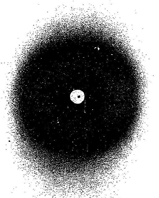
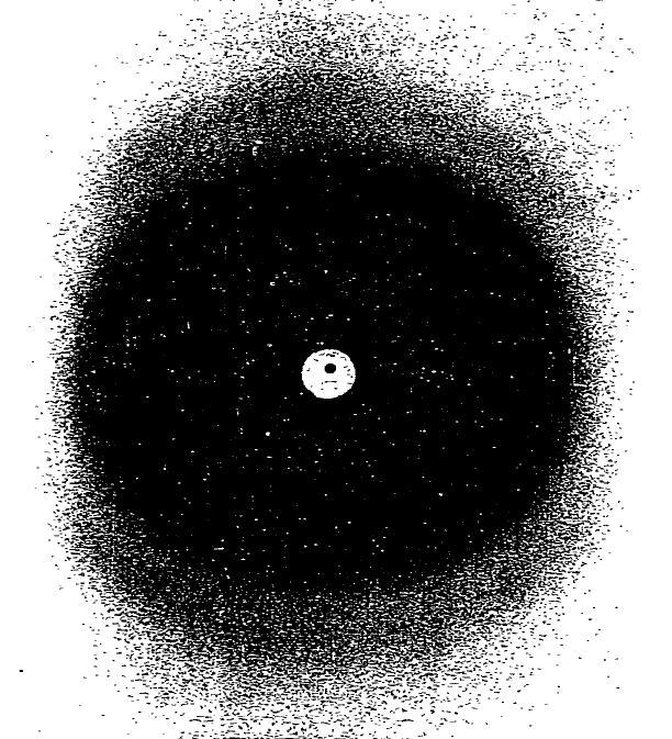
Fig.1 X-Ray fiber diagram of scleroglucan (A) 'as spun' (B) annealed at 45°. *Permission Pending for Diffraction Diagrams
Fig.1 X-Ray fiber diagram of scleroglucan (A) 'as spun' (B) annealed at 45°. *Permission Pending for Diffraction Diagrams
......................................................................................... Scleroglucan Unit Cell Dimensions (a, b, c in Å and α, β, γ in °) (No values are displayed if no information is available or the value is zero) a (Å) = 17.30 - b (Å) = 6.00 - c (Å) = 17.30 - γ (°) = 120.00 Method(s) Used For Structure Determination : X-ray diffraction Link to the Abstract : http://www.sciencedirect.com/science?_ob=ArticleURL&_udi=B6TFF-42H83MY-HG&_user=10&_coverDate=12%2F31%2F1982&_rdoc=1&_fmt=high&_orig=search&_origin=search&_sort=d&_docanchor=&view=c&_searchStrId=1621740018&_rerunOrigin=google&_acct=C000050221&_version=1&_urlVersion=0&_userid=10&md5=34042e26630900e9fb76e5edb62410b6&searchtype=a
Download Structure
|

