![]()
A Database of Polysachharide 3D structures
| Home | |||||||
| About | |||||||
| User Guide | |||||||
| > Amylose A(Revisit-2009) for EXPERTS | |||||||
| Methods | |||||||
| References | |||||||
| Wiki | |||||||
| Contact us | |||||||
|
|
|||||||
Polysaccharides For Experts |
Amylose A(Revisit-2009).........................................................................................
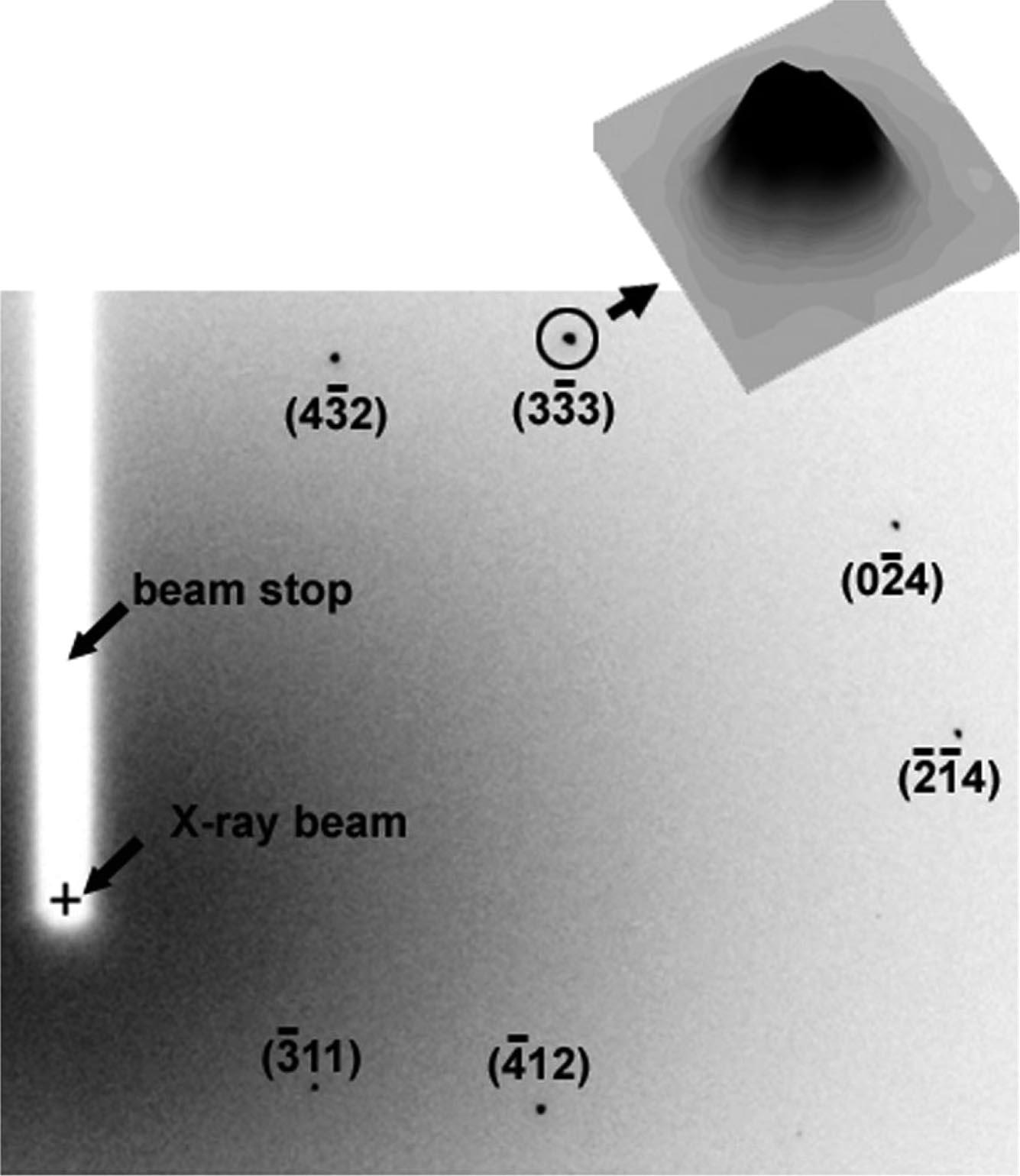
Fig.1 One quadrant of a typical X-ray diffraction diagram of a crystal obtained in 1 s during a 2° rotation. Inset: example of the intensity distribution across one of the diffraction spots (circled). *Permission Pending for Diffraction Diagrams
......................................................................................... Amylose A(Revisit-2009) Unit Cell Dimensions (a, b, c in Å and α, β, γ in °) (No values are displayed if no information is available or the value is zero) a (Å) = 20.83 - b (Å) = 11.45 - c (Å) = 10.58 - γ (°) = 122.00 Link to the Abstract : http://pubs.acs.org/doi/abs/10.1021/ma801789j
Download Structure
Unit A-amylose_2009-popov_expanded
|