![]()
A Database of Polysachharide 3D structures
| Home | |||||||
| About | |||||||
| User Guide | |||||||
| > Agarose (single) for EXPERTS | |||||||
| Methods | |||||||
| References | |||||||
| Wiki | |||||||
| Contact us | |||||||
|
|
|||||||
Polysaccharides For Experts |
Agarose (single).........................................................................................
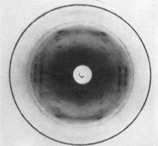
Fig.1 X-ray diffraction patterns from WHT film of agarose. (a)Oriented under second type of stretching procedure between 80 and 100°C. Patterns show high degree of orientation and crystallization with a layer line spacing of 3.552 nm and a clearly defined meridional on the fourth layer line at spacing 0.888 nm. *Permission Pending for Diffraction Diagrams
Fig.1 X-ray diffraction patterns from WHT film of agarose. (a)Oriented under second type of stretching procedure between 80 and 100°C. Patterns show high degree of orientation and crystallization with a layer line spacing of 3.552 nm and a clearly defined meridional on the fourth layer line at spacing 0.888 nm.
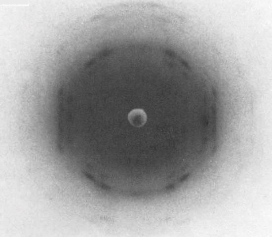
Fig.2 X-ray diffraction patterns from DMAA high-temperature (DHT) film. (a)Oriented using type 2 stretching procedure at 42-53°C. Substantially improved crystallinity showing layer lines at spacing 2.919 nm with a meridional on the third layer line at spacing 0.973 nm. *Permission Pending for Diffraction Diagrams
Fig.1 X-ray diffraction patterns from WHT film of agarose. (a)Oriented under second type of stretching procedure between 80 and 100°C. Patterns show high degree of orientation and crystallization with a layer line spacing of 3.552 nm and a clearly defined meridional on the fourth layer line at spacing 0.888 nm.
Fig.2 X-ray diffraction patterns from DMAA high-temperature (DHT) film. (a)Oriented using type 2 stretching procedure at 42-53°C. Substantially improved crystallinity showing layer lines at spacing 2.919 nm with a meridional on the third layer line at spacing 0.973 nm. (b)Similar to (b) but with slightly different layer line spacing of 2.815 nm and changes in the intensity of various reflections, especially on equator. *Permission Pending for Diffraction Diagrams
Fig.1 X-ray diffraction patterns from WHT film of agarose. (a)Oriented under second type of stretching procedure between 80 and 100°C. Patterns show high degree of orientation and crystallization with a layer line spacing of 3.552 nm and a clearly defined meridional on the fourth layer line at spacing 0.888 nm.
Fig.2 X-ray diffraction patterns from DMAA high-temperature (DHT) film. (a)Oriented using type 2 stretching procedure at 42-53°C. Substantially improved crystallinity showing layer lines at spacing 2.919 nm with a meridional on the third layer line at spacing 0.973 nm. (b)Similar to (b) but with slightly different layer line spacing of 2.815 nm and changes in the intensity of various reflections, especially on equator. (c)Oriented using type 2 stretching procedure at 80-100°C. Pattern is highly crystalline, with layer line spacing 3.552 nm and meridional on fourth layer line. Similar to Fig. (A). *Permission Pending for Diffraction Diagrams
......................................................................................... Agarose (single) Unit Cell Dimensions (a, b, c in Å and α, β, γ in °) (No values are displayed if no information is available or the value is zero) Helix Type : extended and single agarose chains Method(s) Used For Structure Determination : X-ray diffraction Link to the Abstract : http://onlinelibrary.wiley.com/doi/10.1002/bip.360280802/abstract
Download Structure
|